facebook NEUROPATHIES - Pass The OT

NEUROPATHIES


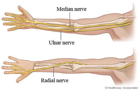
The main nerve entrapments in the upper extremity involve the median nerve, ulnar nerve, or radial nerve. The involved nerve itself may be compressed at two different levels resulting in a double crush syndrome.

As the nerves that control the upper extremity pass through the arm towards the hand, they pass through relatively fixed anatomical structures, or tunnels, usually as the nerve passes a joint. These tunnels are unable to accommodate swelling, therefore when swelling occurs within the limited volume of the tunnel, the nerve is compressed.


Common Compressive Neuropathies

Radial nerve
• Radial tunnel syndrome
• Posterior interosseous syndrome
• Superficial radial nerve syndrome

Ulnar nerve
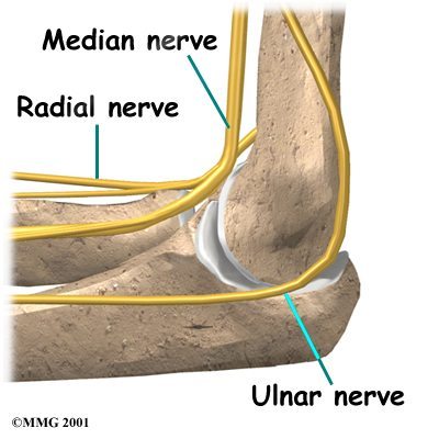
• Cubital tunnel syndrome
• Ulnar tunnel syndrome
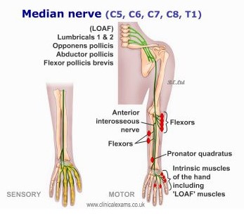
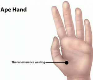
Median nerve
• Pronator syndrome
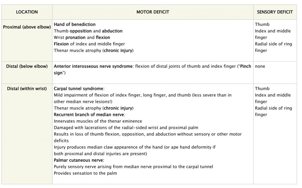
• Anterior interosseous syndrome
• Carpal tunnel syndrome

Although these differ in the anatomic distribution of their symptoms, they share a similar pathophysiology and treatment.
For example: Cubital and Radial Tunnels
Both pass near the elbow and run through the forearm to several fingers. The cubital tunnel houses the ulnar nerve, which winds behind and inside the elbow; the radial tunnel houses the radial nerve on the outside of the elbow. Due to the proximity of these nerves with the skin, they can be irritated by direct pressure – such as by leaning too much on the elbows or bending the arms awkwardly while asleep. 


Radial Nerve                                           

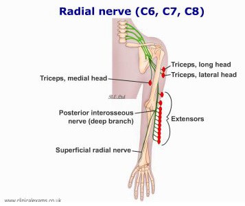
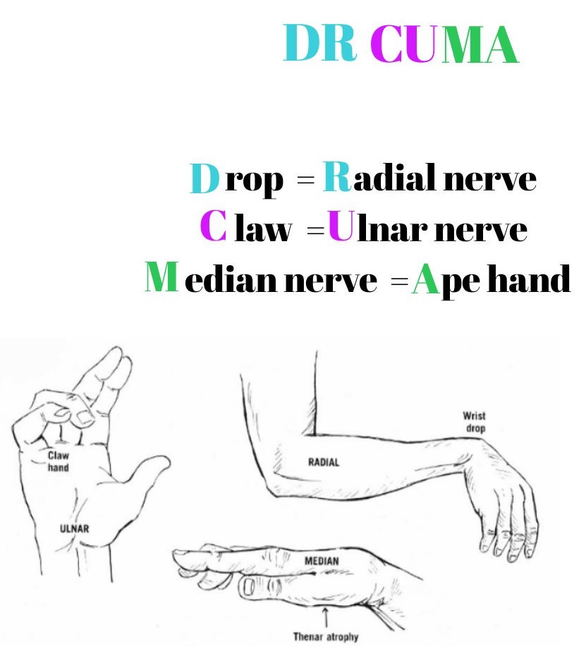
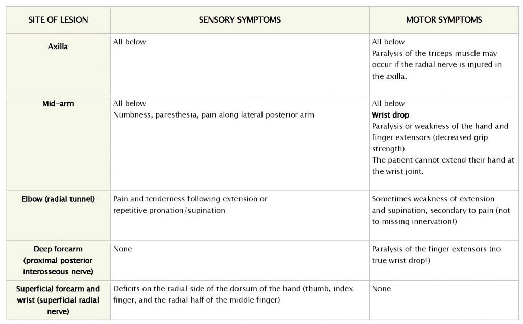
Radial neuropathies are caused by acute or chronic injury to the radial nerve. Clinical presentations vary depending on the mechanism, site, and extent of nerve injury. Radial neuropathies are characterized by sensory symptoms of pain, paresthesia, and numbness, as well as motor symptoms of weakness of extension at the elbow, wrist (“wrist drop”), and/or fingers.

Location and typical causes of injury

• Axilla: Improper crutch use.

• Mid-arm:
–  Fracture of humerus (radial nerve runs in spiral groove).
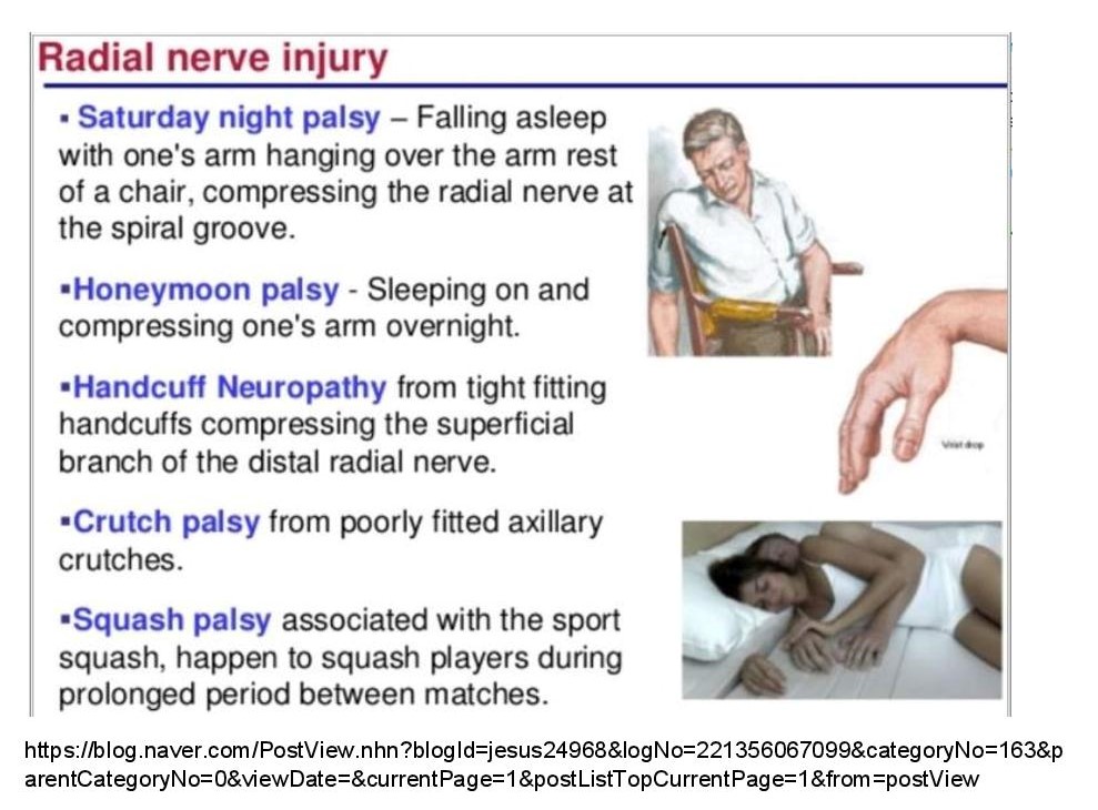
–  Compression of the radial nerve due to draping of arm over furniture (Saturday night palsy) or pressure on mid-arm; more likely in the setting of intoxication, anesthesia, or sleep (honeymoon palsy).

• Elbow: Radial tunnel syndrome due to chronic compression within the radial tunnel.

• Deep forearm: Posterior interosseous nerve syndrome (a syndrome characterized by weakness of muscles in the extensor compartment of the forearm) due to a fracture of the radial head or chronic soft tissue compression.

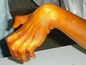
• Superficial forearm or wrist: Superficial radial nerve compression, called “cheiralgia paresthetica,” often due to tight wristwatches or handcuffs or repetitive pronation and supination.
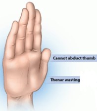
Ulnar nerve                                     

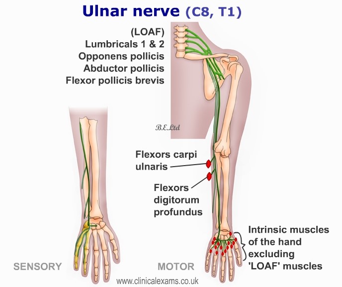
The ulnar nerve is most commonly compressed at or near the cubital tunnel of the elbow and Guyon canal of the wrist. The compression causes paresthesia, numbness, and/or pain in the ulnar nerve distribution. Depending on the site of compression, the patient may experience weakness in certain hand muscles. Ulnar entrapment neuropathy may be suspected based on clinical symptoms and signs, but it must be confirmed by electromyography (EMG).

Clinical Features:

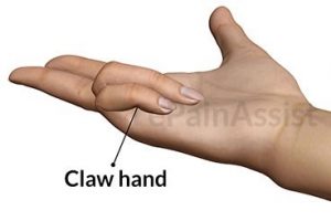
MOTOR– Muscle weakness and atrophy.
Proximal as well as distal lesions lead to claw hand deformity.

SENSORY– Sensory loss over the hypothenar eminence, ulnar 1 ½ fingers.
Lesion at the elbow: Typically presents with referred pain in the forearm. Positive Tinel test → Marked paresthesia can be reproduced in the ulnar portion of the hand by tapping on the medial epicondyle of the humerus. Tinel sign is elicited by lightly percussing along the course of the affected nerve from distal to proximal.
Lesion at the wrist: Sensory symptoms may or may not be present.

Median Nerve                                   

Motor and sensory deficits depend on whether the median nerve lesion is proximal (above the elbow) or distal (below the elbow). While proximal lesions present with the “hand of benediction,” distal lesions present with either the “OK sign” (anterior interosseous nerve syndrome) or, in the case of carpal tunnel syndrome, with mildly impaired thumb and index finger motion. Both proximal lesions and carpal tunnel syndrome result in reduced sensation in the area of the thumb, index and middle finger. Anterior interosseous nerve syndrome does not cause any sensory deficits. Chronic injuries to the nerve result in atrophy of median nerve innervated muscles while acute injuries do not have this feature. Treatment is mostly conservative and focuses on rest and immobilization.


.

Comparison of Compressive Neuropathies

Syndrome Radial Tunnel 


Posterior Interosseous Nerve  Ulnar Tunnel 
AKA- handlebar palsy
Cubital Tunnel Carpal Tunnel
Nerve Radial

Posterior interosseous nerve (PIN)

Radial nerve divides into its 2 terminal branches, the superficial radial nerve and the posterior interosseous nerve. 

Radial

Posterior interosseous nerve (PIN)

Ulnar 


Ulnar  Median
Causes Radial tunnel syndrome and posterior interosseous nerve syndrome are often used interchangeably as both have the same compressive points. They are, however, separate entities.

The most common place for compression of the radial nerve is at the elbow where the nerve enters a tight tunnel made by muscle , bone, and tendon.

Common etiology:
• Microtrauma from repetitive pronation- supination movements
• Trauma- fracture/ dislocation
• Space occupying lesions- e.g. ganglion, lipomas
• Inflammation- e.g. rheumatoid synovitis

Etiology similar to that of radial tunnel syndrome.

• Trauma
• Synovitis (rheumatoid)
• Tumors
• Iatrogenic injuries

Caused by direct compression in Guyon’s canal.

Commonly due to:
• Soft tissue tumor, usually a ganglion which originates in the wrist joint
• Repetitive trauma which can result from the use of a jackhammer
• Chronic pressure which can occur in the hand of a cyclist from the handlebars

The ulnar nerve is especially vulnerable to compression at the elbow because it must travel through a narrow space with very little soft tissue to protect it. When bending your elbow, the ulnar nerve must stretch around the bony ridge of the medial epicondyle. Keeping your elbow flexed for long periods or repeatedly bending your elbow can therefore irritate the nerve.

Commonly caused by:
• Sleeping with your elbow in a flexed position
• Leaning on your elbow for long periods of time
• Fluid buildup in the elbow can cause swelling that may compress the nerve
• A direct blow to the inside of the elbow can cause pain, electric shock sensation, and numbness in the little and ring fingers. This is commonly called “hitting your funny bone.

The carpal tunnel becomes narrowed or the synovium surrounding the flexor tendons swell, putting pressure on the median nerve. Most cases of carpal tunnel syndrome are caused by a combination of factors, such as:
• Repetitive hand use. Repeating the same hand and wrist motions or activities over a prolonged period of time
• Hand and wrist position. Doing activities that involve extreme flexion or extension of the hand and wrist for a prolonged period of time
• Pregnancy. Hormonal changes during pregnancy can cause swelling.
• Health conditions. Diabetes, rheumatoid arthritis, and thyroid gland imbalance are conditions that are associated with carpal tunnel syndrome
Symptoms Associated with lateral elbow and dorsal forearm pain may radiate to the wrist and dorsum of the fingers. Motor dysfunction is not a feature

• Pain only- Deep aching pain in proximal forearm, from lateral elbow to wrist which increases during forearm rotation and lifting activities


• No motor or sensory dysfunction- Muscle weakness due to pain and not muscle denervation


Always associated with motor weakness.

The clinical presentation is characterized by the loss of function due to variable degrees of weakness involving ulnar deviation.


• No sensory deficit. Pain may be present, but it usually is not a primary symptom
• Paresis or paralysis of the digital and thumb extensor muscles, resulting in an inability to extend the thumb and fingers at their metacarpophalangeal joints and weak thumb abduction.

Wrist drop is absent since the extensor carpi radialis is spared, but because of weakness of the extensor carpi ulnaris, there is usually radial deviation of the wrist during extension

Presentation varies based on location of compression within Guyon’s canal and may be:
• Motor only
– weakness of the intrinsic muscles of the hand innervated by the ulnar nerve, which may present as a weakening of the hand grip and clawing of the fourth and fifth digits
– weakness of the adductor pollicis muscle, inability to adduct the thumb
.
• Sensory only
pain and/or paresthesia of medial palm and ulnar half of the fourth digit and entire anterior side of the fifth digit.)
.
• Mixed Motor & Sensory
• Numbness and paresthesia in the
distribution of the ulnar nerve (the small finger and ulnar half of the ring finger
• Weakness of the intrinsic hand muscles
• Rarely have pain
Night-time symptoms are very common because many people sleep with their wrists bent, symptoms may awaken you from sleep.

During the day, symptoms often occur when holding something for a prolonged period of time with the wrist bent forward or backward, such as when using a phone, driving, or reading a book.

Symptoms include:
• Numbness, tingling, burning, and pain primarily in the thumb and index, middle, and ring fingers
• Occasional shock-like sensations that radiate to the thumb and index, middle, and ring fingers
• Pain or tingling that may travel up the forearm toward the shoulder
• Weakness in hand
• Dropping objects due weakness, numbness, or a loss of proprioception


Radial Nerve  Ulnar Nerve Median Nerve
Tests Extension of elbow, wrist and fingers Wartenberg sign: Little finger in persistent abduction due to weak third palmar interosseous muscle.

Wartenberg’s Sign refers to the slightly greater abduction of the fifth digit, due to paralysis of the abducting palmar interosseous muscle and unopposed action of the radial innervated extensor muscles (digiti minimi, digitorum communis ). This should not be confused with Wartenberg’s Syndrome which is described as the entrapment of the superficial branch of the radial nerve with only sensory manifestations and no motor deficits. 

Froment sign:  The thumb flexes at the interphalangeal joint while pinching a piece

of paper to compensate for a weak adductor pollicis muscle.


OK sign: Inability make an “O.K.” sign, as flexion of the IP joint of the thumb and the distal IP joint of the index finger are impaired.
Anterior interosseous nerve is a motor branch of the median nerve.
Phalen test: Both Phalen test and Reverse Phalen’s tests are considered positive when the patient’s symptoms are reproduced, test gives the same symptoms as that experienced with carpal tunnel syndrome, namely paresthesia (buring, tingling, numbness) in the distribution of the median nerve.
Videos

Carpal tunnel syndrome- Tinel’s sign and Phalen’s sign

OK sign

Hand Deformities Wrist drop Claw hand deformity
Partial/Incomplete Claw hand- Involves only ulnar 2 digits as in isolated ulnar nerve palsy.
Ape hand deformity

Hand of Benediction

Pictures


Mnemonic for Nerve Lesions 


Combined Median and Ulnar Nerve Compression: Complete Claw hand involving all digits 

Caused by imbalance between strong extrinsics and deficient intrinsics and characterized by MCP hyperextension, PIP & DIP flexion


Review of Claw Hand, Ape Hand, and the Sign of Benediction

Hand deformity Ulnar claw


Hand of Benediction Ape hand
Nerve lesion


Ulnar nerve High Median nerve Median nerve
Description Permanent fixed position of the hand at rest Active sign- only occurs when attempt made to make a fist (flex the digits).

When hand relaxed, the clawed appearance goes away.

Default position of the injured hand at rest
Presentation 4th and 5th fingers hyperextended at the MCP joints and hyperflexed at the IP joints Seen when patient is asked to make a fist and digits 4 and 5 (ring and little finger) flex but digits 2 and 3 (index and middle fingers) cannot flex at the MCP or IP joints.


All of the digits become aligned in the same dorsal-ventral plane due to paralysis of the thenar muscles which results in an inability to abduct and oppose the thumb. 
Cause Strong extrinsic muscles lead to unopposed extension of the MCP joints. Weakened intrinsic muscles lead to a loss of MCP flexion and a loss of IP extension. The flexor digitorum profundus and flexor digitorum superficialis muscles remain strong and lead to unopposed flexion of the PIP and DIP joints. Unable to flex digits 2 and 3 (index and middle fingers) due to loss of lateral lumbrical action. Due to unopposed radial nerve action on the finger extensors, digits remain in extension. Results in partial flexion of digits 4 and 5, with other digits remaining in extension. The unopposed action of the adductor pollicis which is innervated by the ulnar nerve, results in the thumb becoming adducted and laterally rotated, and thumb opposition inhibited.


Digits affected Digits 4 and 5 Digits 2 and 3 Digit 1/Thumb


Pictures


QUICK REFERENCE


Radial Nerve


Median Nerve


2 quick methods which can be used to screen the functioning of the three main peripheral nerves of the upper extremity


A. Universal sign language


B. “Rock, Paper, Scissors” .

1. Median Nerve: Rock Position

Pronated full fist     


2. Radial Nerve: Paper Position

Extended wrist and digits with forearm pronated, add supination with open palm 


3. Ulnar Nerve: Scissors Position

4th and 5th fingers MCP and PIP joints are flexed; 2nd and 3rd digits are extended and abducted away from each other; Thumb CMC is adducted and the IP is flexed.


Resources
https://www.amboss.com/us/knowledge/Median_nerve_neuropathy
https://www.chesapeakehand.com/2019/05/17/cubital-and-radial-tunnel-syndrome/
https://www.amboss.com/us/knowledge/Radial_neuropathieshttps://www.handandwristinstitute.com/cubital-tunnel-syndrome-treatment-dallas/
http://www.rimed.org/rimedicaljournal/2013/05/2013-05-37-ortho-nerve.pdfhttps://www.verywellhealth.com/radial-nerve-injury-2488802
https://emedicine.medscape.com/article/1285531-overview#a5
https://www.orthobullets.com/
http://www.rimed.org/rimedicaljournal/2013/05/2013-05-37-ortho-nerve.pdf
https://orthoinfo.aaos.org/
https://www.slideshare.net/tmit2/thumbs-up-thumb-injuries-and-their-managementhttps://www.orfit.com/blog/the-peripheral-nerves-a-quick-assessment-of-nerve-functioning-or-rock-paper-scissors/https://emergencymedicinecases.com/elbow-injuries/
https://www.slideshare.net/tmit2/thumbs-up-thumb-injuries-and-their-management
https://www.orfit.com/blog/the-peripheral-nerves-a-quick-assessment-of-nerve-functioning-or-rock-paper-scissors
/https://emergencymedicinecases.com/elbow-injuries/