| IMAGE | MUSCLE | ORIGIN | INSERTION | ACTION | NERVE |
 |
Pectoralis Major | Medial half of clavicle; sternum; upper six costal cartilages | Lateral lip of intertubercular groove of humerus | horizontal adduction | Medial and lateral pectoral nerves |
 |
Pectoralis Minor | External surfaces of ribs 3-5 | Coracoid process of the scapula | Raises ribs in forced inspiration; draws scapula forward and downward | Medial and lateral pectoral nerves |
 |
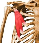
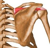
Trapezius | External occipital protuberance; ligamentum nuchae; Spinous processes and surpraspinous ligaments of C7 and all thoracic vertebrae | Lateral third of clavicle; acromion and crest of spine of scapula; Inner/medial aspect of the spine of the scapula | Shrugging the shoulders | Accessory (eleventh cranial) |
 |
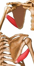
Latissimus Dorsi | Spinous processes of lower six thoracic vertebrae; Lumbar vertebrae, sacral vertebrae, supraspinous ligament; Posterior part of iliac crest; lower three or four ribs; Inferior angle of the scapula | Bottom of intertubercular groove | Extends and hyper- extends the arm, drawing shoulder downward and backward; Extends, adducts and medially rotates the arm | Thoracodorsal (C6-C8) |
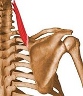 |
Levator Scapulae | Posterior tubercles of transverse processes of C1-C4 | Superior angle of scapula | Elevates scapula (synergist) | Dorsal scapular |
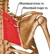 |
Rhomboid Major | Spines of thoracic vertebrae 2-5; Supraspinous ligament | Medial border of the scapula below the spine | Retracts and stabilizes scapula | Dorsal scapular |
| Rhomboid Minor | Spines of c7 and T1; Lower part of ligamentum nuchae | Medial border of the scapula at the root of spine | Retracts and stablizes scapula | Dorsal scapular | |
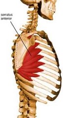 |
Serratus Anterior | Outer surface of first 8-9 ribs | Anterior surface of medial border of scapula | Holds scapula in place against posterior thoracic wall | Long Thoracic |
 |
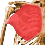
Deltoid | Anterior border and superior surface of lateral third of clavicle; Lateral border of the acromion process; Lower border of the crest of the spine of the scapular | Deltoid tuberosity | Main action: abducts arm; synergist: flexes and medially rotates arm, Extends and laterally rotates arm | Axillary (C5, C6) |
| Rotator Cuff (SITS) | |||||
 |
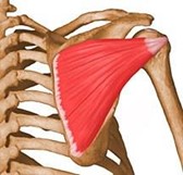
Supraspinatus (rotator cuff) | Supraspinous fossa of scapula | Superior facet of greater tubercle of humerus | Hyperextension of the arm | Suprascapular nerve |
 Infraspinatus (rotator cuff) Infraspinatus (rotator cuff) |
Infraspinous fossa of scapula | Middle facet of the greater tubercle of humerus | Lateral rotation of the arm, synergist to abduction of arm | Suprascapular nerve (C5, C6) | |
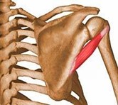 |
Teres Minor (rotator cuff) | Upper 2/3 of the posterior surface of the lateral border of the scapula | Lower facet of the grater tubercle of humerus | Laterally rotates arm (synergist); weak synergist to adduction of arm | Axillary |
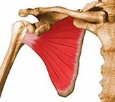 |
Subscapularis (rotator cuff) | Subscapular fossa on the anterior surface of scapula | Lesser tubercle of humerus | Internal rotation of the arm | Upper and lower subscapular nerves (C5, C6) |
 |
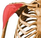
Teres Major | Lower third of the posterior surface of the lateral border of the scapula, near inferior angle | Medial lip of intertubercular groove | Syngergist to latissimus dorsi: Adducts arm, extends arm, medially rotates arm | Lower subscapular nerve |
 |
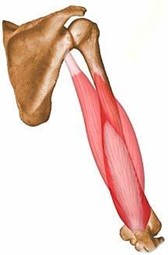
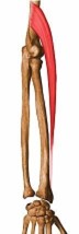
Brachialis | Anterior of lower half of humerus | Coronoid process of ulna; ulnar tuberosity | Primary forearm flexor | Musculocutaneous |
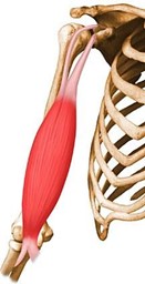 |
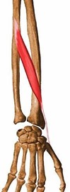
Biceps Brachii | Two heads: Supraglenoid tubercle of scapula & coracoid process of scapula | Radial tuberosity; bicipital aponeurosis into deep fascia on medial side of forearm | Supinates forearm in flexion; weak forearm flexor | Musculocutaneous |
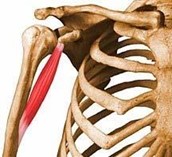 |
Coracobrachialis | Tip of coracoid process of scapula | Middle third of the medial surface of border of humerus | Flexor of humerus at shoulder joint | Musculocutaneous |
 |
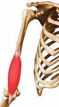
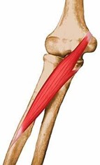
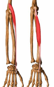
Triceps Brachii | Long head: infraglenoid tubercle of scapula; Lateral head: upper half of posterior surface of humerus; Medial head: posterior surface of lower half of humerus (buried) | Posterior part of olecranon process of ulna | Extends forearm; long head aids in addution if arm is abducted | Radial nerve |
| Superficial Group | |||||
 |
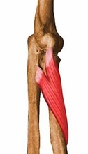
Pronator Teres (superficial group) | Humeral head- medial supracondylar ridge and medial epicondyle of humerus; Ulnar head- medial border of the coronoid process of ulna | Middle of lateral surface of the radius (pronator tuberosity) | Pronates and flexes forearm | Median nerve |
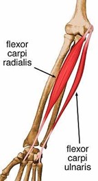 |
Flexor carpi radialus (superficial group) | Medial epicondyle of the humerus through common tendon (flexor pronator tendon) | Front of the bases of the second and third metacarpal bones | Flexes hand; radial deviation | Median nerve |
 |
Palmaris Longus (superficial group) | Medial epicondyle of the humerus through common tendon (flexor pronator tendon) | Front of flexor retinaculum and apex of palmar aponeurosis | Flexes the hand; makes a tight fist | Median nerve |
| Flexor carpi ulnaris (superficial group) | Humeral head- medial epicondyle of humerus; Ulnar head- medial margin of olecranon process of ulna, dorsal border of shaft of ulna | Pisiform bone; hook of hamate; base of fifth metacarpal | Flexes hand; ulnar deviation | Ulnar nerve | |
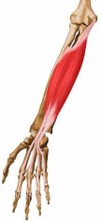 |
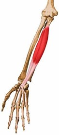
Flexor Digitorum Superficialis (Intermediate grp) | Medial epicondyle of humerus through common tendon; medial margin of coronoid process of ulna; anterior shaft of radius | Four tendons split and connect at middle phalanges of four fingers | Primary: flexion of digits. Flexes carpal region, MCPs & PCPs of digits 2-5. | Median nerve |
|
Flexor Digitorum Profundus (deep) | Upper anterior & medial shaft of ulna and medial coronoid process, interosseous membrane | Front of base of distal phalanges of fingers 2-5 | Flexes distal phalanges (DIPS). Synergist to MCPs & PIPs | Anerior Interosseus/Ulnar (digit flexion backup for median nerve) |
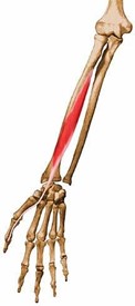 |
Flexor Pollicis Longus | Middle of anterior surface of shaft of radius, interosseous membrane | Palmar aspect of base of distal phalanx of thumb | Flexes the thumb | Anterior interosseous (branch of median) |
| 3 muscles in Anterior Compartment of Forearm- no action on digits: | |||||
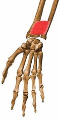 |
Pronator Quadratus | Anterior surface of distal part of shaft of ulna | Lower portion of anterior surface of shaft of radius, distal part of lateral border of radius | Pronates forearm and hand; works with Pronator teres for full pronation. | Anterior interosseous (branch of median) |
 |
Supinator | Lateral epicondyle of humerus; supinator crest of ulna | Posterior and lateral surfaces of upper third of radius | supinates forearm; antagonist of pronator teres and pronator quadratus; supinator when in extension, syngergist in flexion | Radial nerve (because origin off lateral condyle) |
 |
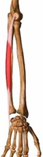
Brachioradialis | Upper 2/3 of lateral supracondylar ridge of humerus | Base of styloid process and lateral surface of radius | Flexes forearm (synergist to brachialis) | Radial nerve (elbow flexion backup) |
| Posterior Compartment of Forearm | |||||
 |
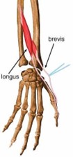
Extensor Carpi Radialis (Brevis3 & Longus2) | Lateral supracondylar ridge and epicondyle of humerus | Dorsal surface of 2nd and 3rd metacarpals | extends hand; synergist in radial deviation with flexor carpi radialis; antagonist to FCR for extension/flexion | Radial |
 |
Extensor Carpi Ulnaris | Common tendon attached to lateral epicondyle of humerus | Dorsal surface of base of 5th metacarpal bone | Extends hand; synergist in ulnar deviation with flexor carpi ulnaris | Radial nerve (because origin off lateral condyle) |
| Extensor Digitorum Communis | Common tendon attached to lateral epicondyle of humerus | Lateral and dorsal surfaces of all phalanges of four fingers | Extends the fingers and wrist; extension expansion joint at distal phalanges | Radial nerve (because origin off lateral condyle) | |
| Extensor Indicis | Posterior surface of ulna and adjacent part of interosseous membrane | Extensor expansion on dorsal surface of proximal phalanx of index finger | Extends index finger | Radial | |
 |
Extensor Digiti Minimi | Common tendon attached to lateral epicondyle of humerus | Dorsal surface of base of first phalanx of fifth finger | Extends fifth finger (pulls out of opposition) | Radial nerve |
 |
Abductor Pollicis Longus | Dorsal surface of shaft or radius, ulna, interosseous membrane | Dorsal surface of base of first metacarpal bone | Abducts thumb & wrist | Radial |
| Extensor Pollicis Brevis | Dorsal surface of radius, adjacent part of interosseous membrane | Base of proximal phalanx of thumb | Extends thumb | Radial | |
 |
Extensor Pollicis Longus | Middle third of dorsal surface of ulna, interosseous membrane | Base of distal phalanx of thumb | Extends thumb | Radial |
| Abductor pollicis Brevis (thenar eminence) | Tubercle of scaphoid, tubercle of trapezium, flexor retinaculum | Lateral aspect of base of proximal phalanx of thumb | Abducts thumb (with Abductor Pollicis Longus) and opposes thumb to other fingers | Median | |
| Flexor Pollicis Brevis (thenar eminence) | Flexor retinaculum and trapezium, first metacarpal bone | Medial and lateral aspect of base of proximal phalanx of thumb | Flexes MCP joint of thumb, opposes thumb | Median/Ulnar | |
| Opponens Pollicis (thenar eminence) | Flexor retinaculum, tubercle of trapezium | Lateral border of first metacarpal bone, wraps around entire lateral border | Rotates thumb into opposition, primary thumb opposer; separates us from animals | Median | |
| Adductor Pollicis | Anterior surface of 2 & 3 metacarpals, capitate and trapezoid | Medial side of proximal phalanx of thumb | adducts thumb | Ulnar | |
| Abductor Digiti Minimi (Hypothenar eminence) | Pisiform bone, tendon of flexor carpi ulnaris | Medial side of proximal phalanx of fifth finger | Abducts fifth finger | Ulnar | |
| Flexor Digiti Minimi (Hypothenar eminence) | Anterior surface of flexor retinaculum, hook of hamate | Medial side of base of proximal phalanx of fifth finger | Flexes fifth finger (extrinisic muscles, Flexor Digitorum Profundus/Superficialis are stronger flexors) | Ulnar | |
| Opponens Digiti Minimi (Hypothenar eminence) | Flexor retinaculum, hook of hamate | Whole length of medial border of fifth metacarpal bone | rotates fifth metacarpal bone, flexing fifth finger | Ulnar | |
| Lumbricales | JUST KNOW: Synergist to PIP & MCP function | ||||
| Palmar Interossei (PAD) | Palmer surface of metacarpals: ADducts digits between 2-3, 3-4, 4-5; | ||||
| Dorsal Interossei (DAB) | Dorsal surface between metacarpals; ABducts between 2-3 and 3-4 digits |
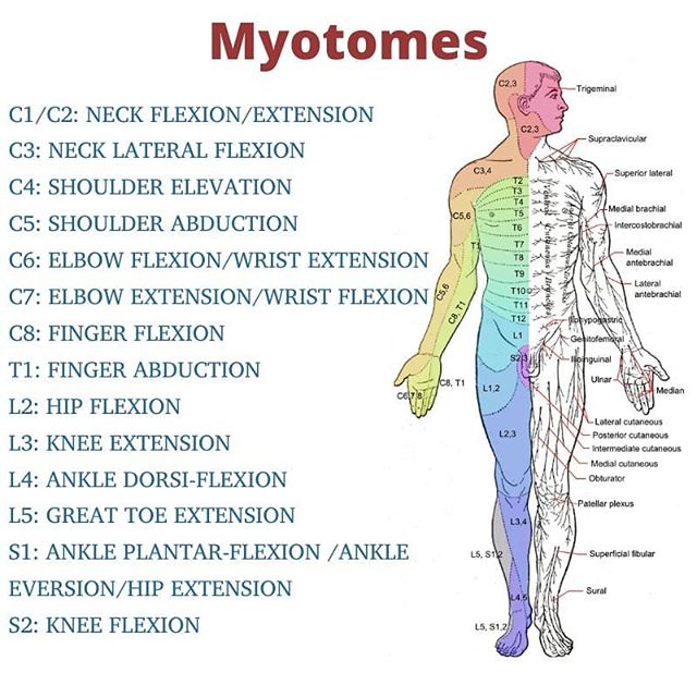
MYOTOMES
The term myotome is defined as a group of muscles innervated by a single spinal nerve root. While a dermatome usually represents a distinct skin area, most roots innervate more than one muscle, and most muscles are innervated by more than one root. For example, the biceps brachii muscle performs flexion at the elbow. It is innervated by the musculocutaneous nerve, which is derived from C5, C6 and C7 nerve roots. Therefore, all three of these spinal nerve roots can be said to be associated with elbow flexion.
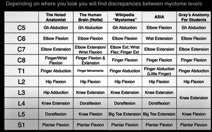
Depending on which resource you refer to, there is a discrepancy between the identified myotomes.
Comparing Myotomes- SCI vs Brachial Plexus Injury
SCI
With a SCI, the loss of motor and/or sensory function is due to damage to neural elements within the spinal canal.
The ASIA Myotome Levels:
The muscles were chosen because of their consistency for being innervated by the segments indicated and their ease of testing in the clinical situation, where testing in any position other than the supine position may be contraindicated.
When using the ASIA, the motor examination is completed through the testing of key muscle functions corresponding to 10 paired myotomes (C5-T1 and L2-S1). It is recommended that each key muscle function should be examined in a rostral-caudal sequence, utilizing standard supine positioning and stabilization of the individual muscles being tested. Improper positioning and stabilization can lead to substitution by other muscles and will not accurately reflect the muscle function being graded.
MYOTOMES
• C5 – Elbow flexion
• C6 – Wrist extension
• C7 – Elbow extension
• C8 – Finger flexion
• T1 – Finger abduction
• L2 – Hip flexion
• L3 – Knee extension
• L4 – Ankle dorsiflexion
• L5 – Great toe extension
• S1 – Ankle plantarflexion
Brachial Plexus Injury
With a brachial plexus lesion/injury it is the peripheral nerves outside the spinal cord that are being affected.
The cervical and thoracic myotomes (C1-T12) are tested with the patient in a seated position. When testing for a brachial plexus lesion, you want to test movement(s) which have the strongest association with each myotome.
Example: The primary muscles involved in the action of arm abduction include the supraspinatus, deltoid, trapezius, and serratus anterior.
• Serratus Anterior, Deltoid- innervated by C5.
• Cranial nerve XI innervates the motor function of the trapezius.
• The supraspinatus muscle is supplied by the suprascapular nerve (C5 and C6)
Technique for assessing C5 myotome with BPL
C5- Shoulder abduction.
Ask the patient to raise both their arms to the side of them simultaneously as strongly as then can while the examiner provides resistance to this movement. Compare the strength of each arm.
MYOTOMES
C5 – Shoulder abduction
C6 – Elbow flexion Wrist extension
C7 – Elbow extension
C8 – Finger flexion
T1 – Finger abduction
L2 – Hip flexion
L3 – Knee extension
L4 – Ankle dorsiflexion
L5 – Great toe extension
S1 – Ankle plantarflexion
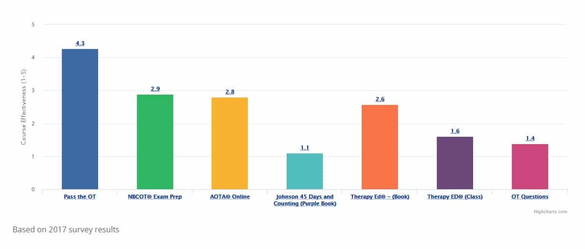
At the end of each month, Pass the OT awards $250 to one lucky user who fills out our brief exit survey.
This survey will be sent to you after you have taken the exam.
We look forward to helping you pass the near future
Begin ProgramPlease upgrade your package to view this quiz
UpgradePlease upgrade your package to view this quiz
UpgradePlease upgrade your package to view full page content
UpgradePlease upgrade your package to view full page content
UpgradeWhat is included
What is not included
What is included
What is not included